• Published on: Jul 30, 2024
  • 4 minute read
  • By: Secondmedic Expert

Ultrasound Imaging Explained: Benefits And Uses In Modern Medicine

  • WhatsApp share link icon
  • copy & share link icon
  • twitter share link icon
  • facebook share link icon

Ultrasound imaging is a key tool in modern medicine, offering a safe and non-invasive way to view the inside of the body. By using high-frequency sound waves, ultrasound creates detailed images without radiation, making it invaluable for diagnosing and monitoring various conditions. In this blog, we’ll explore how ultrasound works, its benefits, and its common uses in healthcare. Join us as we uncover the role of ultrasound in enhancing patient care and advancing medical diagnostics.

What is Ultrasound Imaging?

Ultrasound imaging, also known as sonography, is a medical imaging technique that uses high-frequency sound waves to produce images of the inside of the body. Unlike X-rays or CT scans, ultrasound does not involve radiation, making it a safer option for various diagnostic procedures. This technology is widely used to examine organs, tissues, and blood flow, and it plays a crucial role in both routine and emergency medical assessments.

How Does Ultrasound Imaging Work?

Ultrasound imaging operates by emitting high-frequency sound waves through a transducer, a handheld device that looks like a small wand. Here’s a step-by-step look at the process:

  • Sound Wave Emission: The transducer sends high-frequency sound waves into the body. These sound waves travel through the body and bounce off different tissues and organs.

  • Echo Reception: The sound waves that reflect off tissues return to the transducer as echoes. The strength and timing of these echoes vary depending on the type of tissue they encounter.

  • Image Formation: The echoes are captured by the transducer and sent to a computer, which processes the data to create real-time images. These images display the internal structures of the body, allowing healthcare providers to view and assess them in detail.

  • Equipment Used: The primary equipment includes the transducer (or probe) and a computer with specialized software to generate and display the images. The transducer is often used with a gel applied to the skin to improve the transmission of sound waves.

By capturing and analyzing these sound wave reflections, ultrasound imaging provides valuable insights into the condition and function of internal organs, guiding diagnosis and treatment decisions.

Benefits of Ultrasound Imaging

  • Non-Invasive and Painless: Ultrasound imaging is a non-invasive procedure, meaning it does not require any incisions or needles. Patients typically experience no discomfort during the exam, making it a stress-free diagnostic tool.

  • No Radiation Exposure: Unlike X-rays or CT scans, ultrasound imaging uses sound waves instead of ionizing radiation. This makes it a safer option for frequent use, particularly in sensitive populations such as pregnant women and children.

  • Real-Time Imaging: Ultrasound provides real-time images, allowing healthcare providers to observe dynamic processes as they occur. This feature is crucial for assessing the function of organs and monitoring the progress of certain conditions.

  • Versatility: Ultrasound is a versatile imaging technique used for a wide range of diagnostic purposes. It can evaluate various body parts, from the abdomen to the heart, and is also effective in guiding certain medical procedures.

Common Uses of Ultrasound Imaging

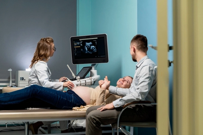
  • Prenatal Care: Ultrasound is widely used in prenatal care to monitor the development of the fetus, check for any abnormalities, and determine the baby’s position and gender. It provides expectant parents with valuable information and reassurance throughout the pregnancy.

  • Cardiology: In cardiology, ultrasound is used to perform echocardiograms, which assess the heart's structure and function. This helps in diagnosing heart diseases, evaluating heart valve function, and guiding treatment decisions.

  • Abdominal and Pelvic Examinations: Ultrasound is used to examine organs in the abdomen and pelvis, such as the liver, kidneys, and bladder. It helps diagnose conditions like kidney stones, liver disease, and bladder abnormalities.

  • Musculoskeletal Imaging: This technique is effective in assessing muscles, tendons, and joints. It is often used to diagnose injuries, such as tears or sprains, and to guide treatment for musculoskeletal conditions.

  • Vascular Studies: Ultrasound is employed to study blood flow and detect issues in blood vessels, such as blockages or clots. This helps in managing conditions like deep vein thrombosis and assessing vascular health.

Ultrasound imaging’s broad applications and significant benefits make it an indispensable tool in modern medicine.

Advancements in Ultrasound Technology

Recent advancements in ultrasound technology have significantly enhanced its diagnostic capabilities and patient experience. Here are some key innovations:

  • 3D and 4D Imaging: Modern ultrasound machines now offer 3D and 4D imaging capabilities, allowing for more detailed and dynamic views of internal structures. 3D imaging provides static, three-dimensional images, while 4D imaging adds the element of movement, which is particularly useful in prenatal care.

  • Doppler Ultrasound: This technology measures and visualizes blood flow within vessels and the heart. It helps in diagnosing conditions related to blood circulation, such as blockages or abnormalities in blood flow.

  • Portable Ultrasound Devices: Advances in technology have led to the development of portable ultrasound machines. These compact devices bring ultrasound capabilities to various settings, including remote or emergency locations, and enable quicker, on-the-go assessments.

  • Elastography: This technique assesses the stiffness of tissues, which can be crucial for diagnosing liver disease or tumors. Elastography provides additional information beyond traditional imaging, aiding in more accurate diagnoses.

  • Artificial Intelligence (AI): AI algorithms are increasingly integrated into ultrasound technology to enhance image quality, automate measurements, and assist in the interpretation of results. AI can improve diagnostic accuracy and streamline the imaging process.

These advancements make ultrasound imaging more versatile, accurate, and accessible, benefiting both patients and healthcare providers.

How to Prepare for an Ultrasound Examination

Preparation for an ultrasound examination can vary depending on the type of exam being performed. Here are some general tips to help ensure a smooth process:

  • Follow Specific Instructions: Your healthcare provider will give you specific instructions based on the type of ultrasound. For instance, you might need to fast for a period before an abdominal ultrasound or drink plenty of water before a pelvic exam.

  • Wear Comfortable Clothing: Choose clothing that allows easy access to the area being examined. For abdominal or pelvic ultrasounds, you may need to wear loose-fitting clothing.

  • Avoid Certain Foods or Drinks: If instructed to fast, avoid eating or drinking anything except water for the specified time. This helps ensure that your stomach and intestines are clear for the examination.

  • Arrive on Time: Arrive at your appointment a few minutes early to complete any necessary paperwork and to ensure that you’re prepared for the exam.

  • Inform the Technician: Let the ultrasound technician know if you have any medical conditions or if you are pregnant, as this may affect the imaging process.

Following these preparation tips will help ensure that your ultrasound examination is conducted smoothly and that the results are accurate.

Conclusion

Ultrasound imaging is a vital tool in modern healthcare, offering a non-invasive and radiation-free method for examining the body's internal structures. Its ability to provide real-time, detailed images makes it indispensable for diagnosing and monitoring a wide range of conditions, from prenatal assessments to cardiac evaluations. With recent advancements in technology, including 3D imaging and portable devices, ultrasound continues to enhance diagnostic accuracy and patient care.

By understanding and utilizing ultrasound imaging, healthcare providers can deliver more precise diagnoses and better manage patient treatment plans. This innovative technology not only supports effective medical decision-making but also contributes to improved patient outcomes and overall health management.

If you have any questions about ultrasound imaging or need to schedule an appointment, don’t hesitate to reach out to us at SecondMedic Healthcare. Our experienced team is here to provide you with the highest quality care and answer any inquiries you may have. Contact us today to learn more or to book your ultrasound examination!

Read FAQs


A. Ultrasound imaging, also known as sonography, uses high-frequency sound waves to create images of the inside of the body. A handheld device called a transducer emits these sound waves, which bounce off tissues and return as echoes. These echoes are then processed by a computer to produce real-time images of internal organs and structures.

A. Yes, ultrasound imaging is considered very safe. It does not use ionizing radiation, unlike X-rays or CT scans, making it a non-invasive and radiation-free diagnostic tool. It is commonly used in prenatal care to monitor the development of fetuses, demonstrating its safety and effectiveness.

A. During an ultrasound examination, a gel will be applied to your skin to help the transducer make secure contact and produce clear images. The technician will move the transducer over the area being examined. The process is typically painless and non-invasive. Depending on the type of exam, you might need to follow specific preparation instructions provided by your healthcare provider.

A. Ultrasound imaging is used for various diagnostic purposes, including prenatal care, cardiology, abdominal and pelvic examinations, musculoskeletal imaging, and vascular studies. It helps in monitoring fetal development, evaluating heart function, diagnosing abdominal and pelvic conditions, assessing musculoskeletal injuries, and examining blood flow in vessels.

Read Blog
Remote Cardiac Monitoring India: Transforming Heart Health Through Continuous Digital Tracking

Remote Cardiac Monitoring India: Transforming Heart Health Through Continuous Digital Tracking

Heart disease remains one of the leading causes of death in India. According to the Indian Council of Medical Research (ICMR), cardiovascular diseases account for nearly 28% of total deaths in the country. With rising stress levels, sedentary lifestyles, air pollution exposure, diabetes, hypertension, and delayed diagnosis, the need for better cardiac monitoring has grown significantly. Remote cardiac monitoring India is emerging as a powerful solution to monitor heart health in real time and detect abnormalities before they turn into emergencies.

Remote cardiac monitoring involves wearable or portable ECG devices that track heart signals continuously or at regular intervals. These devices transmit data securely to cardiologists or AI-based systems, enabling early detection of arrhythmias, rhythm abnormalities, ischemic patterns, and fluctuations in heart rate variability (HRV). SecondMedic brings cutting-edge remote monitoring options that help patients manage heart-related risks effectively, anytime and anywhere.

Why Remote Cardiac Monitoring Is Becoming Essential in India

India has a disproportionately high cardiac burden. Reports by WHO show that Indians suffer heart attacks at a younger age compared to global averages. Additionally, more than 50% of cardiac patients in India do not recognize early warning signs and reach hospitals late.

Remote cardiac monitoring helps solve these challenges by offering:

  • Early arrhythmia detection
     

  • Timely identification of heart stress
     

  • Post-surgery cardiac recovery supervision
     

  • Reduced hospital dependency
     

  • Continuous heart rhythm tracking
     

  • Better outcomes for chronic heart patients
     

With easy access to digital ECG devices, monitoring is no longer limited to hospitals.

How Remote Cardiac Monitoring Works

Remote cardiac monitoring uses smart ECG patches, chest straps, handheld ECG devices, and even AI-driven wearables. These devices record heart electrical activity or rhythm patterns and transmit the data to:

  • Cardiologists
     

  • Digital dashboards
     

  • AI algorithms
     

  • Care teams
     

The real-time nature of the data allows immediate response in case of abnormalities.

Core parameters captured include:

  • ECG waveform
     

  • Heart rate variability (HRV)
     

  • Arrhythmias (AFib, SVT, PVCs)
     

  • Tachycardia or bradycardia
     

  • QT interval abnormalities
     

  • Stress-related heart rhythm changes
     

SecondMedic ensures that the captured data is clinically relevant and accessible to both patients and doctors.

Patients Who Benefit Most from Remote Cardiac Monitoring

1. Post-Cardiac Surgery Patients

After angioplasty, bypass surgery, pacemaker implantation, or stent placement, patients require strict monitoring for weeks or months. Remote devices track recovery trends.

2. Individuals With Arrhythmia

Conditions like atrial fibrillation (AFib) require continuous monitoring, as episodes may come and go unpredictably.

3. Patients With Heart Failure

Monitoring helps evaluate worsening symptoms or cardiac stress.

4. High-Risk Individuals

Patients with diabetes, hypertension, obesity, or family history of heart disease benefit from early detection.

5. Elderly and Homebound Patients

Remote monitoring eliminates frequent hospital visits for routine ECG checks.

6. People Experiencing Palpitations

Intermittent symptoms can be captured and analyzed with wearable monitors.

Key Advantages of Remote Cardiac Monitoring

Early Detection Saves Lives

Abnormal rhythms such as atrial fibrillation can go unnoticed and lead to stroke. Remote monitoring detects these in real time.

Avoids Emergency Situations

AI alerts allow doctors to intervene early before symptoms worsen.

Better Long-Term Cardiac Care

Continuous data provides better insights than occasional clinic-based ECGs.

High Accuracy

Medical-grade ECG wearables match hospital-level precision for diagnosis.

Patient Convenience

Patients can monitor their heart health from home, office, or while traveling.

Improved Doctor Decisions

Trend-based cardiac data supports more accurate treatment planning.

Role of AI in Remote Cardiac Monitoring

AI and machine learning models analyze patterns collected from ECG devices.

AI enhances care by:

  • Identifying subtle abnormalities
     

  • Predicting arrhythmia events
     

  • Detecting ischemic trends early
     

  • Classifying ECG signal types
     

  • Reducing manual interpretation burden
     

SecondMedic integrates AI-assisted analysis to strengthen cardiac risk evaluation.

Post-Surgery and Rehabilitation Monitoring

Cardiac rehabilitation is crucial after major procedures. Remote monitoring provides:

  • Heart rate control
     

  • Blood pressure trend tracking
     

  • Rhythm abnormality detection
     

  • Activity monitoring
     

  • Medication adherence support
     

This reduces readmission rates and supports safer recovery.

Remote Cardiac Monitoring in Rural India

Many small towns lack cardiology specialists or advanced ECG equipment. Remote monitoring bridges this gap by:

  • Connecting patients to cardiologists virtually
     

  • Providing ECG analysis without hospital visits
     

  • Reducing delay in diagnosis
     

  • Supporting long-term monitoring at home
     

SecondMedic helps democratize heart care access across India.

Challenges and Solutions

Device Accuracy Variability

Solution: Use medical-grade, certified ECG wearables.

Digital Literacy Issues

Solution: Simple interfaces, guided onboarding, multilingual instructions.

Data Security

Solution: End-to-end encrypted data under DPDP Act compliance.

Connectivity Challenges

Solution: Devices with offline sync capabilities.

Future of Remote Cardiac Monitoring in India

The next decade will bring significant advancements:

  • Patch-based continuous ECG devices
     

  • AI-driven early warning scores
     

  • Smart pacemaker data integration
     

  • Multi-day continuous ECG monitoring
     

  • Predictive arrhythmia detection algorithms
     

  • Tele-ICU support for remote patients
     

SecondMedic is actively working to integrate next-generation cardiac monitoring tools into its platform.

Conclusion

Remote cardiac monitoring India is transforming heart care by enabling real-time ECG tracking, early abnormality detection, and continuous cardiologist oversight. This digital-first approach empowers high-risk patients, improves outcomes, and ensures safer long-term cardiac management. SecondMedic’s integrated digital heart monitoring solutions allow patients across India to access expert cardiac care anytime.

To explore remote cardiac monitoring solutions, visit www.secondmedic.com


References

  • ICMR - Cardiovascular disease burden
     

  • WHO - Early cardiac risk detection
     

  • NITI Aayog - Digital health innovation in India
     

  • ABDM - Unified digital health mission
     

  • Statista - Wearable and ECG device adoption India
     

  • SecondMedic cardiac care insights

See all

Live Doctor consultation
Live Doctor Chat

Download Our App & Get Consultation from anywhere.

App Download
call icon for mobile number calling and whatsapp at secondmedic